
CHEEKS · ILLUSTRATED 3D RECOVERY
Zygomatic Arch Reduction (Side Width): before & after and recovery stages
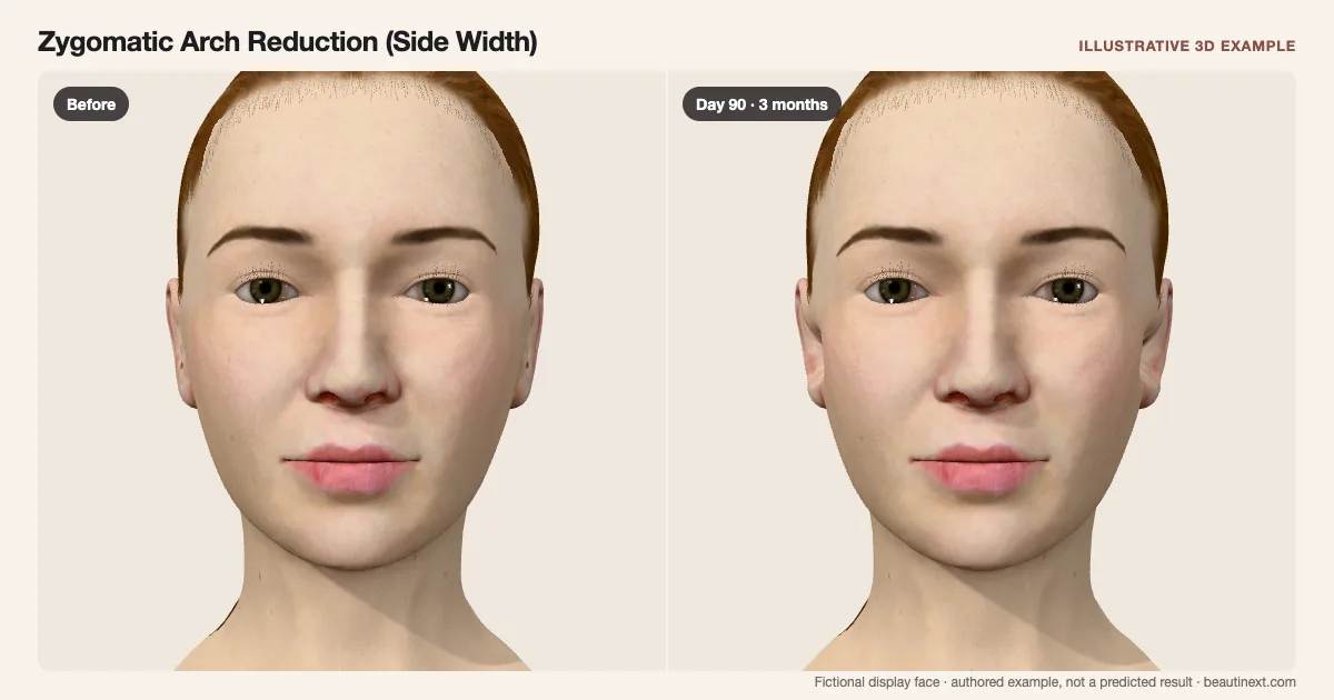
Compare the width in front of the ears as early swelling gives way to a narrower side outline.
SUMMARY
What this example shows
The side of the face in front of the ears narrows as an example of arch repositioning, while the front cheekbone body, lower cheeks and jaw are retained. Early fullness settles separately. Osteotomy design, fixation, temporal hollowing and bone-to-skin ratios are not calculated.
STAGE BY STAGE
Zygomatic Arch Reduction (Side Width) recovery stages
Before treatment
The unchanged display face provides a reference for this example.
First few days: Fullness masks the width
Temporary swelling in front of the ears overlaps the narrower outline.
Postoperative swelling is described in combined facial-contouring reports.
Around 2 weeks: Early settling
Surface color fades while fullness remains.
These dates illustrate a course; reports do not set individual milestones.
Around 1 month: Side comparison
Less fullness reveals the narrower arch region.
Bone movement cannot directly predict the visible skin change.
Around 3 months: Later width example
The side outline is easier to compare while the front cheekbone stays fixed.
Soft-tissue response and temporal hollowing need separate assessment.
TIMELINE
Day-by-day checkpoints in the 3D model
The simulator interpolates between these authored checkpoints. They describe the illustration, not measured clinical averages.
The unchanged display face provides a reference for this example.
Swelling overlaps the narrowed side of the face.
Temporary fullness in front of the ears partly conceals the new width.
Local swelling begins easing in this example.
Less color reveals more of the side outline.
The arch region is narrower with residual fullness.
Little early fullness remains on the surface.
The example does not predict sagging, temporal hollowing or bone union.
INDIVIDUAL DIFFERENCES
How recovery can vary
Timing and response
This narrows only the side of the face in front of the ears. The front cheekbone body, lower-cheek hollow and jaw are retained.
Longer term
Osteotomy design and fixation affect settling. The final frame does not predict sagging, symmetry or bone union.
EVIDENCE NOTES
Sources and limitations
- Journal of Craniofacial Surgery: Intraoral zygoma reduction using L-shaped osteotomy · 2014
Describes body and arch osteotomies and facial-width planning in reduction malarplasty. It does not measure the visible skin change or set recovery dates.
- Journal of Craniofacial Surgery: Malar arch movement study
A CT study of inward malar movement. The authors call for further soft-tissue analysis; bone measurements do not calibrate this surface animation.
- Annals of Translational Medicine: Combined facial contouring case series
Postoperative swelling in a small combined jaw/malar surgery series. It cannot establish an isolated-procedure or individual recovery clock.
KEEP COMPARING
Other cheeks examples
This illustration is not medical advice, diagnosis, a treatment recommendation or a prediction of personal results. Swelling, color, contour and dates are authored examples on a fictional face. BeautiNext is not a clinic; your treating team’s assessment and written recovery plan take priority.