
EARS · ILLUSTRATED 3D RECOVERY
Ear Reduction, Upper Ear (Macrotia): before & after and recovery stages
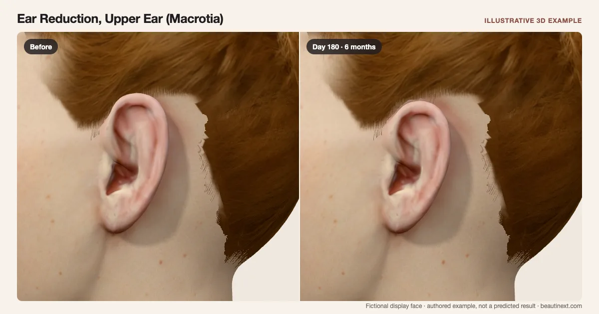
Compare an enlarged upper ear with its reduced outline as early fullness and a residual line change.
SUMMARY
What this example shows
A fixed enlarged upper ear becomes shorter and narrower while the lobe and attachment stay in place. Temporary swelling and color settle around the reduced outline, and an illustrative line inside the upper rim fades but remains. This upper-ear example does not pin the ear backward, reconstruct missing cartilage or calculate surgical dimensions.
STAGE BY STAGE
Ear Reduction, Upper Ear (Macrotia) recovery stages
Enlarged upper-ear reference
The same fictional upper ear is taller and wider; the lobe is unchanged.
This starting shape is not a diagnosis attached to a regional face.
First few days: Reduction under swelling
A shorter upper outline is partly hidden by swelling and color; a line sits inside the rim.
The view omits dressings to show the surface. The line is one illustrative location, not an incision plan.
Around 1 month: Outline and remaining line
Less fullness reveals the shorter, narrower upper ear and a colored line.
General ear surgery recovery varies; one month does not establish comfort or scar maturity.
Around 6 months: Later upper-ear comparison
The reduced outline remains and the illustrative line is faint but still present.
Scars and symmetry can differ or continue changing beyond this comparison point.
TIMELINE
Day-by-day checkpoints in the 3D model
The simulator interpolates between these authored checkpoints. They describe the illustration, not measured clinical averages.
The unchanged display face provides a reference for this example.
The smaller outline is partly obscured by local fullness and color.
Temporary swelling overlays the upper-ear reduction.
Some local fullness and a visible closure line remain.
The upper rim is easier to compare as the early response eases.
The upper ear is shorter and narrower; a colored line remains.
The illustrated line fades while the reduced outline remains.
A faint line remains. This date is not a personal healing deadline.
INDIVIDUAL DIFFERENCES
How recovery can vary
Timing and response
This depicts upper-ear size reduction, with the lobe and attachment fixed. Combined pinning, lobe reduction, missing-ear reconstruction, dressings and internal cartilage surgery are not reconstructed.
Longer term
Dates, dimensions and line fading are authored examples. A calmer surface does not establish sensation, cartilage flexibility, scar maturity or a date for pressure on the ears.
EVIDENCE NOTES
Sources and limitations
- Cleveland Clinic: Ear reduction and general recovery
Distinguishes size reduction from pinning and describes incisions, swelling and variable recovery. It does not calibrate this upper-ear display.
- Walid Sabbagh · Ear Reconstruction: Upper-ear reduction approach
A specialist describes upper-ear height and width reduction with a scar inside the helix. This technique-specific account is not a universal recovery or scar forecast.
KEEP COMPARING
Other ears examples
This illustration is not medical advice, diagnosis, a treatment recommendation or a prediction of personal results. Swelling, color, contour and dates are authored examples on a fictional face. BeautiNext is not a clinic; your treating team’s assessment and written recovery plan take priority.