
EYELIDS · ILLUSTRATED 3D RECOVERY
Buried-Suture Double Eyelid Surgery: before & after and recovery stages
Follow small puncture marks and temporary puffiness as a buried-suture fold becomes easier to see.
SUMMARY
What this example shows
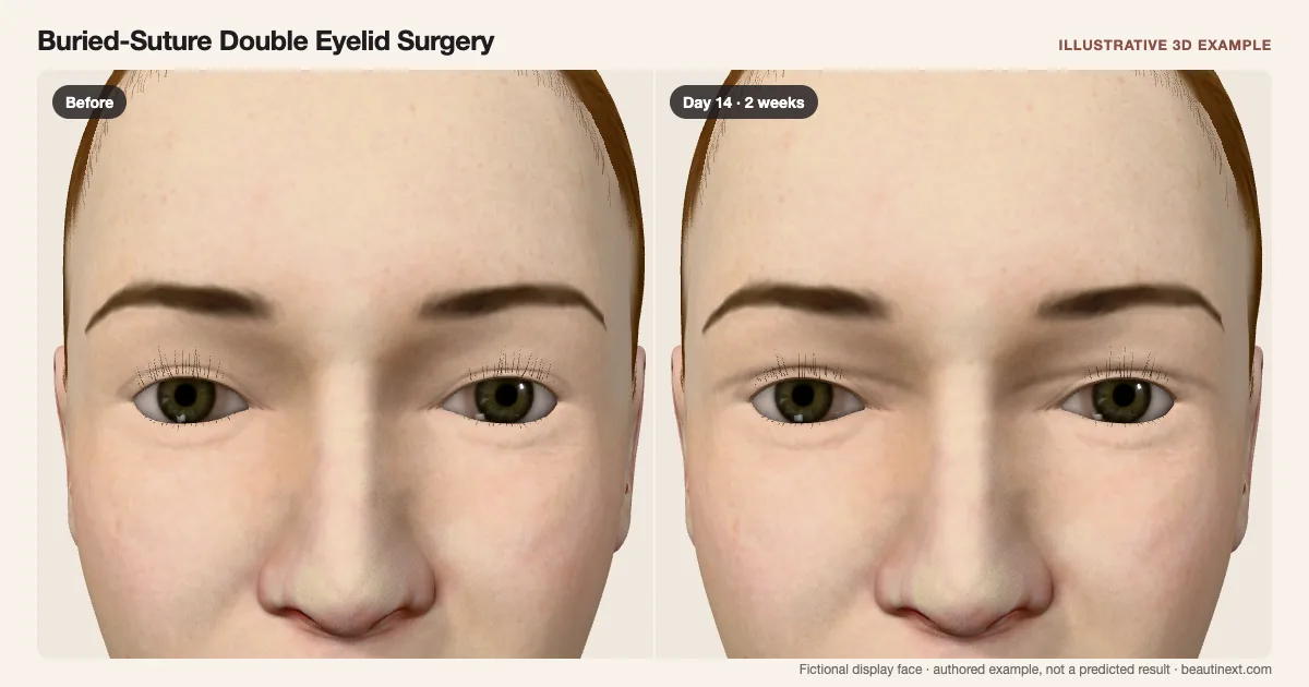
Buried sutures support an upper-lid fold through small puncture sites. The example shows three early marks per lid and temporary fullness, without a continuous skin incision or skin removal. Existing folds remain part of the display face. The line, puncture positions and dates are illustrative; no specific knot pattern, crease height or durability is predicted.
STAGE BY STAGE
Buried-Suture Double Eyelid Surgery recovery stages
Before treatment
The unchanged display face provides a reference for this example.
First day: Small marks and fullness
Three small marks per lid sit along the illustrated fold.
One puncture-based approach is shown. There is no continuous skin incision or external stitch row.
Around 1 week: Marks becoming quieter
The puncture marks fade while some fullness remains.
Fading surface marks do not mean buried sutures have been removed.
Around 2 weeks: Fold easier to compare
The example marks are no longer visible; the supported fold remains.
These dates illustrate one course, not a recovery deadline or proof of internal healing.
Around 3 months: Later retained-fold example
The illustration retains the crease after the early surface response.
Creases can loosen or disappear. This endpoint does not promise lasting retention.
TIMELINE
Day-by-day checkpoints in the 3D model
The simulator interpolates between these authored checkpoints. They describe the illustration, not measured clinical averages.
The unchanged display face provides a reference for this example.
Local marks and fullness surround the supported fold.
Small marks become quieter while temporary fullness remains.
The skin marks fade; buried support is not shown being removed.
The crease remains as the illustrated surface response eases.
The example fold remains without a continuous incision scar.
Maintaining this example crease is not a promise of permanent retention.
INDIVIDUAL DIFFERENCES
How recovery can vary
Timing and response
This is a buried-suture example. It does not remove excess skin or simulate ptosis repair. Existing folds stay part of the chosen face; appearance does not determine suitability for this method.
Longer term
The fold may change beyond this view. Internal sutures, individual scarring and the timing or likelihood of crease loss are not reconstructed.
EVIDENCE NOTES
Sources and limitations
- SNEC · HealthXchange: Double-eyelid methods
Distinguishes puncture-based suture fold creation from small-incision and open techniques. Describes shorter recovery and possible loss of the fold; it does not establish this model's dates or severity.
- AAO EyeWiki: Suture and incisional crease techniques
Buried fixation, technique variation and possible loosening or loss of a crease. The surface model does not reproduce internal knots or predict durability.
KEEP COMPARING
Other eyelids examples
- Subbrow Lift (Upper-Lid Skin)
- Upper Blepharoplasty
- Eyelid Revision (Lowering a High Crease)
- Eyelid Revision (Asymmetric Crease)
- Double Eyelid Surgery
- Partial-Incision Double Eyelid Surgery
- Ptosis Repair (Upper-Lid Height)
- Epicanthal Fold Restoration
- Epicanthoplasty (Inner Corner)
- Lateral Canthopexy (Lower-Lid Support)
- Lateral Canthoplasty (Outer Corner)
- Lower-Lid Fat Repositioning
- Lower Blepharoplasty
- Under-Eye Fat Transfer
- Tear-Trough Filler
- Crow’s Feet Botox (Anti-Wrinkle Injections)
This illustration is not medical advice, diagnosis, a treatment recommendation or a prediction of personal results. Swelling, color, contour and dates are authored examples on a fictional face. BeautiNext is not a clinic; your treating team’s assessment and written recovery plan take priority.