
EYELIDS · ILLUSTRATED 3D RECOVERY
Lateral Canthoplasty (Outer Corner): before & after and recovery stages
Follow the outer corner and adjacent lower lid as early fullness recedes.
SUMMARY
What this example shows
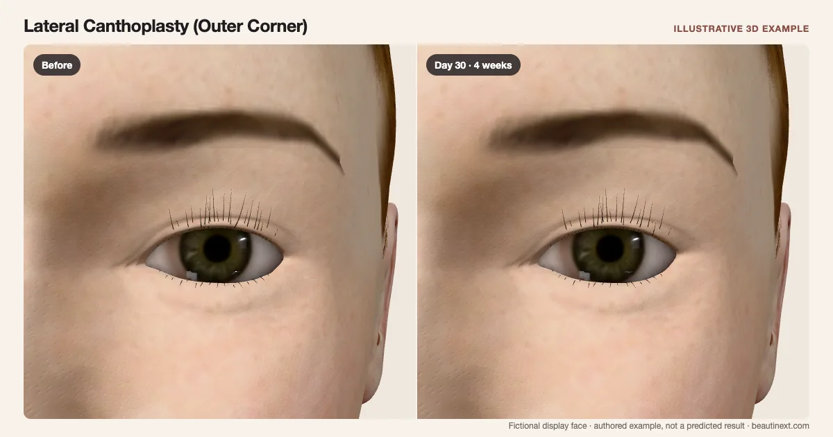
The outer lid junction is repositioned upward with adjacent lower-lid support. The central upper-lid margin and inner corner are retained. This is an outer-corner reconstruction example, not inner-corner surgery or a canthopexy technique simulation.
STAGE BY STAGE
Lateral Canthoplasty (Outer Corner) recovery stages
Before treatment
The unchanged display face provides a reference for this example.
First few days: Outer-corner fullness
Local swelling masks the supported outline.
Early swelling and bruising can make the eyelids look uneven.
Around 2 weeks: Outline emerging
Less surface color exposes the outside lid junction.
The visible early response often improves over the first weeks.
Around 1 month: Corner position clearer
The outer corner remains raised in this example.
A clearer outline does not measure tendon or eye-surface recovery.
Around 3 months: Later corner example
The illustration shows little early fullness.
Incision lines can remain visible for several months.
TIMELINE
Day-by-day checkpoints in the 3D model
The simulator interpolates between these authored checkpoints. They describe the illustration, not measured clinical averages.
The unchanged display face provides a reference for this example.
Local fullness surrounds the changed outer lid position.
Swelling and color obscure the supported outline.
The outside lid junction becomes easier to compare.
The visible reaction becomes lighter in this example.
The outer corner remains raised after the early response.
Incision lines can keep fading after visible swelling improves.
INDIVIDUAL DIFFERENCES
How recovery can vary
Timing and response
This outer-corner reconstruction example does not represent inner-corner surgery, every canthopexy technique or a chosen amount of eye widening.
Longer term
Eye closure, comfort and scars are not shown by the corner position alone. The final checkpoint is not a guarantee of symmetry or a return-to-activity date.
EVIDENCE NOTES
Sources and limitations
- Cleveland Clinic: Canthoplasty
Outer-corner reconstruction, early swelling/bruising and incision lines fading over months.
KEEP COMPARING
Other eyelids examples
- Subbrow Lift (Upper-Lid Skin)
- Upper Blepharoplasty
- Eyelid Revision (Lowering a High Crease)
- Eyelid Revision (Asymmetric Crease)
- Double Eyelid Surgery
- Partial-Incision Double Eyelid Surgery
- Buried-Suture Double Eyelid Surgery
- Ptosis Repair (Upper-Lid Height)
- Epicanthal Fold Restoration
- Epicanthoplasty (Inner Corner)
- Lateral Canthopexy (Lower-Lid Support)
- Lower-Lid Fat Repositioning
- Lower Blepharoplasty
- Under-Eye Fat Transfer
- Tear-Trough Filler
- Crow’s Feet Botox (Anti-Wrinkle Injections)
This illustration is not medical advice, diagnosis, a treatment recommendation or a prediction of personal results. Swelling, color, contour and dates are authored examples on a fictional face. BeautiNext is not a clinic; your treating team’s assessment and written recovery plan take priority.