
SKIN & SCARS · ILLUSTRATED 3D RECOVERY
Mole Removal (Excision): before & after and recovery stages
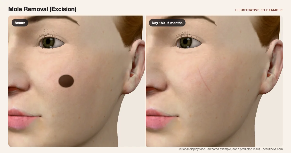
Compare the same fictional cheek mole with a sutured line and a later residual linear scar.
SUMMARY
What this example shows
A closed line and early stitches replace the same raised reference. One fictional cheek mole is used for comparison. Relief is shaded on the surface; open wounds and dressings are omitted. This does not diagnose a mole or select a removal method.
STAGE BY STAGE
Mole Removal (Excision) recovery stages
Raised brown reference
The same fixed cheek spot supplies the starting view.
Appearance alone does not establish whether a mole is harmless.
Early example: Closed line and stitches
Three small crossing stitches sit on a longer closed line.
The number and direction are illustrative; dressings and deeper tissue are omitted.
Around 3 weeks: Closed scar line
Stitches are no longer shown; the colored line remains.
The authored day-8 stitch change is not a removal schedule.
Around 6 months: Residual linear scar
A faint line remains instead of the round starting spot.
Width, color and maturation vary; dates do not predict an individual scar.
TIMELINE
Day-by-day checkpoints in the 3D model
The simulator interpolates between these authored checkpoints. They describe the illustration, not measured clinical averages.
The same fixed cheek spot supplies the starting view.
A local mark fades while residual scarring remains.
A local mark fades while residual scarring remains.
A local mark fades while residual scarring remains.
A local mark fades while residual scarring remains.
A local mark fades while residual scarring remains.
A local mark fades while residual scarring remains.
A local mark fades while residual scarring remains.
A local mark fades while residual scarring remains.
A local mark fades while residual scarring remains.
A local mark fades while residual scarring remains.
INDIVIDUAL DIFFERENCES
How recovery can vary
Timing and response
This is one closed-surface excision example, not an incision plan, margin calculation or cancer treatment simulator. A clinician assesses the mole and removed tissue.
Longer term
A scar remains and may change beyond six months. The display does not predict recurrence or further treatment.
EVIDENCE NOTES
Sources and limitations
- AAD: Moles: Diagnosis and treatment
Professional assessment, shave removal or excision with closure, and examination of removed tissue.
- Cleveland Clinic: Mole removal
Shave versus surgical removal, healing over weeks, residual scars and possible recurrence.
KEEP COMPARING
Other skin & scars examples
- Z-Plasty (Scar Reorientation)
- Scar Revision Surgery
- Mole Removal (Shave)
- Microdermabrasion
- Dermabrasion
- Light Chemical Peel
- Medium Chemical Peel
- Deep Chemical Peel
- Ablative Laser Resurfacing
- Nonablative Laser Resurfacing
- Microneedling (Without RF)
- Radiofrequency Microneedling
- IPL (Brown-Spot Response)
- Laser Toning (Melasma)
- Skin Booster Injections
- TCA CROSS (Ice-Pick Scars)
- Punch Elevation (Boxcar Scars)
- Punch Skin Grafting (Acne Scars)
- Punch Excision (Acne Scars)
- Cryotherapy (Small Raised Scars)
- Keloid Excision
- Steroid Injections (Keloid)
- Steroid Injections (Raised Acne Scars)
- Subcision (Rolling Cheek Scars)
- Pulsed Dye Laser (Cheek Redness)
This illustration is not medical advice, diagnosis, a treatment recommendation or a prediction of personal results. Swelling, color, contour and dates are authored examples on a fictional face. BeautiNext is not a clinic; your treating team’s assessment and written recovery plan take priority.