
SKIN & SCARS · ILLUSTRATED 3D RECOVERY
Mole Removal (Shave): before & after and recovery stages
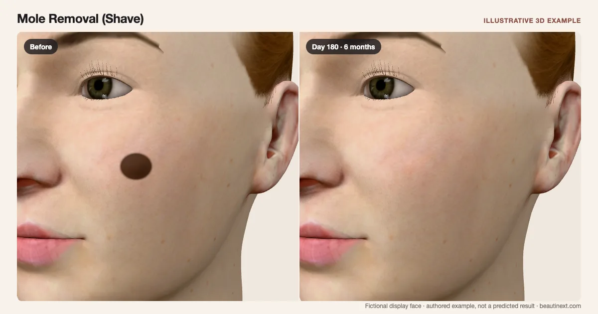
Compare one fictional raised cheek mole with a round surface mark that fades but remains visible.
SUMMARY
What this example shows
A round surface mark remains after the raised reference disappears. One fictional cheek mole is used for comparison. Relief is shaded on the surface; open wounds and dressings are omitted. This does not diagnose a mole or select a removal method.
STAGE BY STAGE
Mole Removal (Shave) recovery stages
Raised brown reference
One fixed cheek spot has a raised appearance.
A fictional display mark, not a diagnosis of the selected face.
Early example: Round local mark
The brown raised reference is gone; local color and surface residue remain.
Open wounds and dressings are omitted from this surface illustration.
Around 3 weeks: Quieter surface
The round site is flatter and less colored in this example.
Healing varies; this date is a comparison point.
Around 6 months: Residual round scar
A faint round difference remains at the original site.
The example does not promise invisible scarring.
TIMELINE
Day-by-day checkpoints in the 3D model
The simulator interpolates between these authored checkpoints. They describe the illustration, not measured clinical averages.
One fixed cheek spot has a raised appearance.
A local mark fades while residual scarring remains.
A local mark fades while residual scarring remains.
A local mark fades while residual scarring remains.
A local mark fades while residual scarring remains.
A local mark fades while residual scarring remains.
A local mark fades while residual scarring remains.
A local mark fades while residual scarring remains.
A local mark fades while residual scarring remains.
A local mark fades while residual scarring remains.
A local mark fades while residual scarring remains.
INDIVIDUAL DIFFERENCES
How recovery can vary
Timing and response
Mole assessment and removal method require a clinician. Size, depth, site and healing change the appearance; this example omits dressings and open wounds.
Longer term
Scars or pigment changes can persist. Recurrence is possible and needs reassessment; it is not animated here.
EVIDENCE NOTES
Sources and limitations
- AAD: Moles: Diagnosis and treatment
Professional assessment, shave removal or excision with closure, and examination of removed tissue.
- Cleveland Clinic: Mole removal
Shave versus surgical removal, healing over weeks, residual scars and possible recurrence.
- West Suffolk NHS: Curettage, cautery and shave excision
Small-lesion shave procedures and a local scar whose color can change over months. This is general leaflet guidance, not a calibration of the model.
KEEP COMPARING
Other skin & scars examples
- Z-Plasty (Scar Reorientation)
- Scar Revision Surgery
- Mole Removal (Excision)
- Microdermabrasion
- Dermabrasion
- Light Chemical Peel
- Medium Chemical Peel
- Deep Chemical Peel
- Ablative Laser Resurfacing
- Nonablative Laser Resurfacing
- Microneedling (Without RF)
- Radiofrequency Microneedling
- IPL (Brown-Spot Response)
- Laser Toning (Melasma)
- Skin Booster Injections
- TCA CROSS (Ice-Pick Scars)
- Punch Elevation (Boxcar Scars)
- Punch Skin Grafting (Acne Scars)
- Punch Excision (Acne Scars)
- Cryotherapy (Small Raised Scars)
- Keloid Excision
- Steroid Injections (Keloid)
- Steroid Injections (Raised Acne Scars)
- Subcision (Rolling Cheek Scars)
- Pulsed Dye Laser (Cheek Redness)
This illustration is not medical advice, diagnosis, a treatment recommendation or a prediction of personal results. Swelling, color, contour and dates are authored examples on a fictional face. BeautiNext is not a clinic; your treating team’s assessment and written recovery plan take priority.